Supracondylar Fracture: Image 1, Normal Annotated

Averyl Shindruk, MD & Michael Schick, DO
Full description
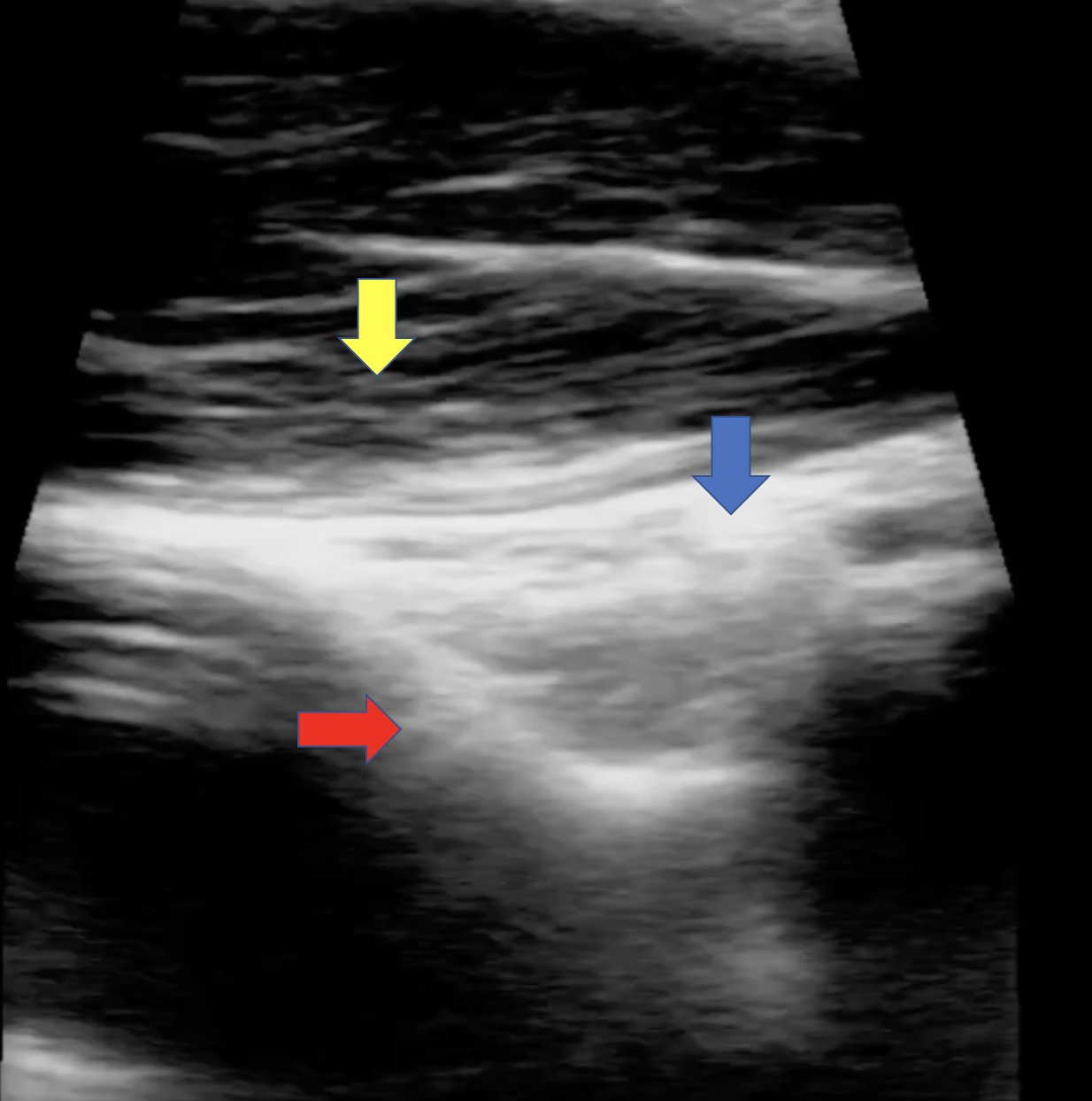
Non-injured distal arm for comparison. Ultrasound image of normal long axis of the distal arm with intact cortex (red arrow), adjacent overlying tendon (yellow arrow) without separation by fluid, fat pad (blue arrow) is flat and does not bulge above humeral line.
Download image “Supracondylar Fracture: Image 1, Normal Annotated ”
- typeImage
- created on
- file formatpng
- file size655 KB